Taxonomic group: bacteria / Proteobacteria
(Phylum: Proteobacteria)
NCBI PubMed ID: 32512087Publication DOI: 10.1016/j.ijbiomac.2020.06.019Journal NLM ID: 7909578Publisher: Butterworth-Heinemann
Correspondence: christoph.hundschell

tu-berlin.de
Institutions: Technical University Berlin, Faculty III Process Sciences, Institute of Food Technology and Food Chemistry, Department of Food Technology and Food Material Science, Straße des 17. Juni 135, 10623 Berlin, Germany, Technical University of Munich, Chair of Technical Microbiology, Gregor-Mendel-Straße 4, 85354 Freising, Germany
Levan is a bacterial homopolysaccharide, which consists of β-2→6 linked β-(D)-fructose monomers. Because of its structural properties and its health promoting effects, levan is a promising functional ingredient for food, cosmetic and pharmaceutical products. The properties of levan have been reported to be linked to its molecular weight. For a better understanding of how the molecular weight determines its polymer conformation in aqueous solution, levan produced by the food grade acetic acid bacterium Gluconobacter albidus TMW 2.1191 was analysed over a broad molecular weight range using dynamic and static light scattering and viscometry. Low molecular weight levan exhibit a compact random coil structure. As the molecular weight increases, the structure transforms into a compact non-drained sphere. The density of the sphere continues to increase with increasing molecular weight. This resulted in a negative exponent in the Mark-Houwink-Sakurada Plot. For the first time, an increase in molecular density with increasing molecular weight, as determined by a negative Mark-Houwink-Sakurada exponent, can be shown for biopolymers. Our results reveal the unique properties of high-molecular weight levan and confirm the need of further systematic studies on the structure-function relationship of levan for its targeted use in food, cosmetic and pharmaceutical applications.
polysaccharide conformation, Dynamic light scattering, intrinsic viscosity, Mark-Houwink-Sakurada, Static light scattering
Structure type: oligomer ; 10000-10000000000
Location inside paper: p.399, Fig.1
Trivial name: levan
Compound class: EPS
Contained glycoepitopes: IEDB_142488,IEDB_146664,IEDB_923066,IEDB_983931,SB_192
Methods: conformation analysis, viscometry, MALLS, photometry, DLS
NCBI Taxonomy refs (TaxIDs): 318683Reference(s) to other database(s): GTC:G87428FH
Show glycosyltransferases
There is only one chemically distinct structure:
Taxonomic group: fungi / Ascomycota
(Phylum: Ascomycota)
Organ / tissue: mycelium
The structure was elucidated in this paperNCBI PubMed ID: 31047067Publication DOI: 10.1016/j.carbpol.2019.03.104Journal NLM ID: 8307156Publisher: Elsevier
Correspondence: He L <kite006
126.com>
Institutions: Institute of Biochemistry, College of Life Sciences, Zhejiang University, Hangzhou, China, Key Laboratory of State Forest Food Resources Utilization and Quality Control, Zhejiang Provincial Key Laboratory of Forest Food, Zhejiang Academy of Forestry, Hangzhou, China, Department of Processing, Marine Fisheries Research Institute of Zhejiang, Zhoushan, China, Department of Food Science, Rutgers University, New Brunswick, NJ, USA
The fine structure and chain conformation of a heteropolysaccharide (PCIPS3) from mycelium of Paecilomyces cicadae were investigated via the analysis of HPLC, IR, methylation, NMR spectroscopy and multiangle light scattering. It was determined to be a 22300 g/mol heteropolysaccharide primarily composed of glucose, galactose and mannose in a molar ratio of 23.8:2.1:1.0. The PCIPS3 backbone consisted of 1,4-linked α-D-Glcp and 1,4-linked 6-O-Me-α-D-Glcp residues, which were occasionally interrupted by branched β-Galf residues through 1,6-linkage. Moreover, the α (0.60) from Mark-Houwink-Sakurada (MHS) equation suggested that PCIPS3 adopted a flexible chain conformation in 0.1 mol/L NaNO3 at 25 °C. The worm-like chains model parameters for PCIPS3 were estimated as following: ML = 437 nm-1, q = 0.46 nm and 0.79 nm, which were further evidenced by AFM. Furthermore, PCIPS3 showed excellent scavenging capacities of 2,2-diphenyl-1-picrylhydrazyl radical, superoxide radical, hydroxyl radical, ORAC radical and moderate immunomodulatory activity.
structural elucidation, biological activity, heteropolysaccharide, AFM, Paecilomyces cicadae
Structure type: structural motif or average structure ; 22300
Location inside paper: p. 276, Fig. 4, p. 276, Table 3
Compound class: polysaccharide
Contained glycoepitopes: IEDB_130701,IEDB_136044,IEDB_136095,IEDB_137472,IEDB_140629,IEDB_141794,IEDB_142488,IEDB_144983,IEDB_144998,IEDB_146664,IEDB_152206,IEDB_153217,IEDB_190606,IEDB_420417,IEDB_420418,IEDB_420421,IEDB_857742,IEDB_983930,IEDB_983931,SB_165,SB_166,SB_187,SB_192,SB_195,SB_44,SB_67,SB_7,SB_72,SB_88
Methods: 13C NMR, 1H NMR, methylation, NMR-2D, IR, GC-MS, acid hydrolysis, viscosity measurement, extraction, acetylation, methylation analysis, SEC, reduction, CC, cell growth, dialysis, antioxidant activities, cell viability assay, precipitation, phenol-sulphuric acid assay, derivatization, Bradford method, AFM, photometry, HPLC-UV, immunomodulatory activity analysis
Biological activity: polysaccharide showed excellent scavenging capacities of DPPH radical (IC50=0.28 mg/mL), superoxide radical (IC50=0.46 mg/mL), hydroxyl radical (IC50=0.32 mg/mL) and ORAC radical (1623.44 μmol Trolox/g). In addition, it exhibited a moderate immunomodulatory activity by enhancing the secretion of major inflammatory cytokines in macrophages such as TNF-α, IL-1β, IL-6
Comments, role: the published 13C NMR spectrum in DSS was shifted 1.6 ppm upfield by CSDB staff, to accord to a TMS reference
NCBI Taxonomy refs (TaxIDs): 218633
Show glycosyltransferases
NMR conditions: in D2O / DSS at 333 K
[as TSV]
13C NMR data:
Linkage	Residue	C1	C2	C3	C4	C5	C6
6,4	aDGlcp	100.90	72.79	74.59	78.09	72.44	61.68
6,6	Me	55.25
6	aDGlcp	99.82	72.74	74.66	78.09	72.25	67.20
	bDGalf	108.24	82.61	77.64	82.61	76.84	67.20
5,4	aDGlcp	101.04	72.74	74.00	70.58	74.14	62.30
5	aDManp	103.82	?	71.21	77.67	74.28	63.90
5,2	aDGlcp	101.04	72.74	74.00	70.58	74.14	62.30
5	bDGalp	105.53	78.06	72.42	71.24	75.76	?
5,6	aDGlcp	101.04	72.74	74.00	70.58	74.14	62.30
5	bDGlcp	96.95	75.00	76.87	70.58	75.78	67.25


1H NMR data:
Linkage	Residue	H1	H2	H3	H4	H5	H6
6,4	aDGlcp	5.40	3.64	3.97	3.66	3.85	3.85
6,6	Me	3.23
6	aDGlcp	4.97	3.59	4.03	3.66	3.91	3.68
	bDGalf	5.19	4.15	4.11	4.13	3.96	3.68
5,4	aDGlcp	5.40	3.57	3.70	3.43	3.75	3.77-3.86
5	aDManp	5.05	4.13	4.08	3.85	3.77	3.68-3.73
5,2	aDGlcp	5.40	3.57	3.70	3.43	3.75	3.77-3.86
5	bDGalp	4.64	3.68	3.79	3.97	3.73	?
5,6	aDGlcp	5.40	3.57	3.70	3.43	3.75	3.77-3.86
5	bDGlcp	4.66	3.28	3.55	3.52	3.68	3.77-4.00


1H/13C HSQC data:
Linkage	Residue	C1/H1	C2/H2	C3/H3	C4/H4	C5/H5	C6/H6
6,4	aDGlcp	100.90/5.40	72.79/3.64	74.59/3.97	78.09/3.66	72.44/3.85	61.68/3.85
6,6	Me	55.25/3.23
6	aDGlcp	99.82/4.97	72.74/3.59	74.66/4.03	78.09/3.66	72.25/3.91	67.20/3.68
	bDGalf	108.24/5.19	82.61/4.15	77.64/4.11	82.61/4.13	76.84/3.96	67.20/3.68
5,4	aDGlcp	101.04/5.40	72.74/3.57	74.00/3.70	70.58/3.43	74.14/3.75	62.30/3.77-3.86
5	aDManp	103.82/5.05	?/4.13	71.21/4.08	77.67/3.85	74.28/3.77	63.90/3.68-3.73
5,2	aDGlcp	101.04/5.40	72.74/3.57	74.00/3.70	70.58/3.43	74.14/3.75	62.30/3.77-3.86
5	bDGalp	105.53/4.64	78.06/3.68	72.42/3.79	71.24/3.97	75.76/3.73	?/?
5,6	aDGlcp	101.04/5.40	72.74/3.57	74.00/3.70	70.58/3.43	74.14/3.75	62.30/3.77-3.86
5	bDGlcp	96.95/4.66	75.00/3.28	76.87/3.55	70.58/3.52	75.78/3.68	67.25/3.77-4.00
1H NMR data:
| Linkage | Residue | H1 | H2 | H3 | H4 | H5 | H6 |
|---|
| 6,4 | aDGlcp | 5.40 | 3.64 | 3.97 | 3.66 | 3.85 | 3.85 |
| 6,6 | Me | 3.23 | |
| 6 | aDGlcp | 4.97 | 3.59 | 4.03 | 3.66 | 3.91 | 3.68 |
| | bDGalf | 5.19 | 4.15 | 4.11 | 4.13 | 3.96 | 3.68 |
| 5,4 | aDGlcp | 5.40 | 3.57 | 3.70 | 3.43 | 3.75 | 3.77
3.86 |
| 5 | aDManp | 5.05 | 4.13 | 4.08 | 3.85 | 3.77 | 3.68
3.73 |
| 5,2 | aDGlcp | 5.40 | 3.57 | 3.70 | 3.43 | 3.75 | 3.77
3.86 |
| 5 | bDGalp | 4.64 | 3.68 | 3.79 | 3.97 | 3.73 | ? |
| 5,6 | aDGlcp | 5.40 | 3.57 | 3.70 | 3.43 | 3.75 | 3.77
3.86 |
| 5 | bDGlcp | 4.66 | 3.28 | 3.55 | 3.52 | 3.68 | 3.77
4.00 |
|
13C NMR data:
| Linkage | Residue | C1 | C2 | C3 | C4 | C5 | C6 |
|---|
| 6,4 | aDGlcp | 100.90 | 72.79 | 74.59 | 78.09 | 72.44 | 61.68 |
| 6,6 | Me | 55.25 | |
| 6 | aDGlcp | 99.82 | 72.74 | 74.66 | 78.09 | 72.25 | 67.20 |
| | bDGalf | 108.24 | 82.61 | 77.64 | 82.61 | 76.84 | 67.20 |
| 5,4 | aDGlcp | 101.04 | 72.74 | 74.00 | 70.58 | 74.14 | 62.30 |
| 5 | aDManp | 103.82 | ? | 71.21 | 77.67 | 74.28 | 63.90 |
| 5,2 | aDGlcp | 101.04 | 72.74 | 74.00 | 70.58 | 74.14 | 62.30 |
| 5 | bDGalp | 105.53 | 78.06 | 72.42 | 71.24 | 75.76 | ? |
| 5,6 | aDGlcp | 101.04 | 72.74 | 74.00 | 70.58 | 74.14 | 62.30 |
| 5 | bDGlcp | 96.95 | 75.00 | 76.87 | 70.58 | 75.78 | 67.25 |
|
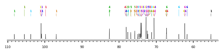
The spectrum also has 2 signals at unknown positions (not plotted). |
There is only one chemically distinct structure:
 report error
 Found 2 records.
Displayed records from 1 to 2
Found 2 records.
Displayed records from 1 to 2
