Taxonomic group: fungi / Basidiomycota
(Phylum: Basidiomycota)
Organ / tissue: mycelium
The structure was elucidated in this paperNCBI PubMed ID: 3723343Publication DOI: 10.1248/yakushi1947.106.3_206Journal NLM ID: 0413613Publisher: Tokyo: Nihon Yakugakkai
Institutions: Tokyo Research Laboratory, Japan Synthetic Rubber Co., Ltd., Japan, Higashi-Yurigaoka 3-5-1, Asao-ku, Kawasaki, Kanagawa, Japan
Structural analysis was made on the glucan moiety of purified antitumor active polysaccharide fraction H11 obtained from the mycelia of P. cocos. In the 13C-nuclear magnetic resonance spectrum of fraction H11, signals at 102.9 and 84.8 ppm were observed, and assigned to C-1 of the β-linkage and C-3 of the β-D-(1,3)-linked D-glucosyl redidues, respectively. On addition of NaOD solution, the C-1 signal was separated into two signals at 104.2 and 104.8 ppm which can be assigned to the C-1 signal of the β-D-(1,3)-linked and the β-D-(1,6)-linked D-glucosyl residues, respectively. The molecular weight of fraction H11 was calculated to be approx. 5*10^6 by the gel filtration method with CPG-1000 Å. Complete methylation and hydrolysis of fraction H11 gave 2,4,6-tri-, 2,3,4-tri-, 2,4-di-, and 2,3,4, 6-tetra-O-methyl glucose as alditol acetates in a molar ratio of 3.96 : 0.95 : 1.05 : 1.00. From these results, it has been revealed that fraction H11 is a highly branched (1,3)-(1,6)-β-D-glucan containing each component in 4 : 1 ratio.
13C-NMR, sarcoma 180, β-D-glucan, Polyporaceae, mycelium, Poria cocos, antitumor effect
Structure type: structural motif or average structure ; 5000000
Location inside paper: H11, Table 1, fig. 2
The structure in this paper was incorrect:
Compound class: glucan
Contained glycoepitopes: IEDB_135614,IEDB_141806,IEDB_142488,IEDB_146664,IEDB_241101,IEDB_983931,SB_192
Methods: 13C NMR, periodate oxidation, GC, Smith degradation, enzymatic digestion, acetylation, methylation analysis, reduction with NaBH4, sulfuric acid hydrolysis, trimethylsilylation, light-scattering
Comments, role: article in Japanese; NMR temperature was not specified; published erroneous NMR chemical shift of #6_bDGlcp C5 (69.2) was removed by CSDB staff; structure was revised by CSDB staff based on the published methylation data
Related record ID(s): 42682
NCBI Taxonomy refs (TaxIDs): 81056
Show glycosyltransferases
NMR conditions: in D2O
[as TSV]
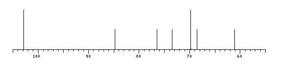
13C NMR data:
Linkage	Residue	C1	C2	C3	C4	C5	C6
3	bDGlcp	102.9	73.5	84.8	68.5	76.5	61.1
6,6	bDGlcp
6	bDGlcp	102.9	?	?	69.7	?	?
	bDGlcp	?	?	?	?	?	69.9


1H NMR data:
missing...
13C NMR data:
| Linkage | Residue | C1 | C2 | C3 | C4 | C5 | C6 |
|---|
| 3 | bDGlcp | 102.9 | 73.5 | 84.8 | 68.5 | 76.5 | 61.1 |
| 6,6 | bDGlcp | |
| 6 | bDGlcp | 102.9 | ? | ? | 69.7 | ? | ? |
| | bDGlcp | ? | ? | ? | ? | ? | 69.9 |
|
The spectrum also has 9 signals at unknown positions (not plotted). |
There is only one chemically distinct structure:
 Found
Found  report error
 (later renamed to: Wolfiporia cocos)