Taxonomic group: plant / Streptophyta
(Phylum: Streptophyta)
Organ / tissue: leaf
The structure was elucidated in this paperNCBI PubMed ID: 9687205Publication DOI: 10.1016/s0008-6215(98)00072-xJournal NLM ID: 0043535Publisher: Elsevier
Institutions: Institute of Chemistry, Slovak Academy of Sciences, Bratislava, Slovak Republic
From the medicinal plant Rudbeckia fulgida, var. sillivantii (Boynton et Beadle) a low-molecular-mass (4-O-methyl-alpha-D-glucurono)-D-xylan was isolated by alkaline extraction, followed by ethanol precipitation, ion-exchange chromatography and gel filtration. The results of compositional and linkage analyses, supported by those of 1H and 13C NMR measurements of oligomers generated on partial acid hydrolysis, showed the (1-->4)-linked beta-D-xylopyranosyl backbone with about 18% of 4-O-methyl-D-glucuronic acid attached to O-2 of the xylose residues. From the mean distance of adjacent carboxyl groups, obtained from experimentally determined single-ion activity coefficients of calcium counterions, it followed that the uronic acid units are separated and distributed regularly along the xylan chain, i.e. approximately each sixth D-xylose unit bears a 4-O-methyl-D-glucuronic acid residue.
Structure determination, Rudbeckia fulgida, (4-O-methyl-alpha-D-glucurono)-D-xylan, H-1 and C-13 NMR spectroscopy
Structure type: structural motif or average structure ; n=18; 17440
Location inside paper: p. 105, Scheme, table 1
Compound class: glucuronoxylan
Contained glycoepitopes: IEDB_114701,IEDB_115136,IEDB_140630,IEDB_167188,IEDB_174332
Methods: 13C NMR, 1H NMR, methylation, IR, GC-MS, extraction, TOCSY, HPGPC, DQF-COSY, HSQC, optical rotation determination, TFA hydrolysis, paper chromatorgraphy
NCBI Taxonomy refs (TaxIDs): 52312Reference(s) to other database(s): GTC:G84935YZ
Show glycosyltransferases
NMR conditions: in D2O at 298 K
[as TSV]
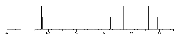
13C NMR data:
Linkage	Residue	C1	C2	C3	C4	C5	C6
4,4	bDXylp	102.6	73.6	74.6	77.2	63.9
4	bDXylp	102.6	73.6	74.6	77.2	63.9
2,4	Me	60.8
2	aDGlcpA	98.5	73.1	?	83.3	72.9	177.5
	bDXylp	102.3	77.7	72.1	76.9	?


1H NMR data:
Linkage	Residue	H1	H2	H3	H4	H5	H6
4,4	bDXylp	4.45	3.26	3.56	3.77	3.37-4.09
4	bDXylp	4.45	3.26	3.56	3.77	3.37-4.09
2,4	Me	3.46
2	aDGlcpA	5.27	3.56	3.74	3.20	4.31	-
	bDXylp	4.61	3.45	3.61	3.79	3.42-4.13


1H/13C HSQC data:
Linkage	Residue	C1/H1	C2/H2	C3/H3	C4/H4	C5/H5	C6/H6
4,4	bDXylp	102.6/4.45	73.6/3.26	74.6/3.56	77.2/3.77	63.9/3.37-4.09
4	bDXylp	102.6/4.45	73.6/3.26	74.6/3.56	77.2/3.77	63.9/3.37-4.09
2,4	Me	60.8/3.46
2	aDGlcpA	98.5/5.27	73.1/3.56	?/3.74	83.3/3.20	72.9/4.31
	bDXylp	102.3/4.61	77.7/3.45	72.1/3.61	76.9/3.79	?/3.42-4.13
1H NMR data:
| Linkage | Residue | H1 | H2 | H3 | H4 | H5 | H6 |
|---|
| 4,4 | bDXylp | 4.45 | 3.26 | 3.56 | 3.77 | 3.37
4.09 | |
| 4 | bDXylp | 4.45 | 3.26 | 3.56 | 3.77 | 3.37
4.09 | |
| 2,4 | Me | 3.46 | |
| 2 | aDGlcpA | 5.27 | 3.56 | 3.74 | 3.20 | 4.31 |
|
| | bDXylp | 4.61 | 3.45 | 3.61 | 3.79 | 3.42
4.13 | |
|
13C NMR data:
| Linkage | Residue | C1 | C2 | C3 | C4 | C5 | C6 |
|---|
| 4,4 | bDXylp | 102.6 | 73.6 | 74.6 | 77.2 | 63.9 | |
| 4 | bDXylp | 102.6 | 73.6 | 74.6 | 77.2 | 63.9 | |
| 2,4 | Me | 60.8 | |
| 2 | aDGlcpA | 98.5 | 73.1 | ? | 83.3 | 72.9 | 177.5 |
| | bDXylp | 102.3 | 77.7 | 72.1 | 76.9 | ? | |
|
The spectrum also has 2 signals at unknown positions (not plotted). |
There is only one chemically distinct structure:
 Found
Found  report error