Taxonomic group: bacteria / Proteobacteria
(Phylum: Proteobacteria)
Associated disease: infection due to Burkholderia pseudomallei [ICD11:
XN3LD 
]
The structure was elucidated in this paperNCBI PubMed ID: 34474230Publication DOI: 10.1016/j.jpba.2021.114340Journal NLM ID: 8309336Publisher: London: Elsevier
Correspondence: (J. Yan <zijie1011

yeah.net>
Institutions: Department of Clinical Microbiology and Immunology, College of Pharmacy and Medical Laboratory, Army Medical University (Third Military Medical University), Chongqing 400038, PR China
Burkholderia pseudomallei causes melioidosis - an infectious disease with high mortality. Its varied clinical manifestations and resistance to many antibiotics make it a potential biothreat agent and calls for a robust diagnostic assay and effective vaccines. Bacterial cell surface polysaccharides are considered a valuable target for diagnostics and as protective antigen candidates. This study characterized the structure of polysaccharides of B. pseudomallei clinical strain from Hainan, China. A novel structural domain [→3-(α-D-Manp-1→3-α-D-Manp)2-2Me-α-L-6dTalp-1→] was identified by chemical analysis, gas chromatography-mass spectrometry (GC-MS), and 1D/2D nuclear magnetic resonance (NMR) spectroscopy. Immunofluorescence and enzyme-linked immunosorbent assay (ELISA) showed that the serum antibodies against the purified polysaccharide antigen could recognize and bind specifically to B. pseudomallei strains. Additionally, the assays revealed cross-reactivity with polysaccharides from different clinical strains. The polysaccharide antigen also exhibited a strong reaction with the sera from melioidosis patients. Thus, the pentasaccharide repeating unit residue could be a potential candidate antigen for the melioidosis serodiagnosis and vaccine development.
polysaccharide, Burkholderia pseudomallei, Structural characterization, immunological characteristics
Structure type: structural motif or average structure
Location inside paper: p. 114340-6, table 3, BPC006-BPPI-b1 fraction
Methods: 13C NMR, 1H NMR, methylation, NMR-2D, GC-MS, ELISA, anion-exchange chromatography, composition analysis, GPC, UV, extraction, statistical analysis, immunization, immunofluorescence analyses
Comments, role: minor β-1,3-linked 6-deoxy-Hepp might be existing separately; NMR temperature was not specified.
Related record ID(s): 10446, 10447, 10979
NCBI Taxonomy refs (TaxIDs): 1229785
Show glycosyltransferases
NMR conditions: in D2O
[as TSV]
13C NMR data:
Linkage	Residue	C1	C2	C3	C4	C5	C6	C7
2	Ac	?	19.10
	bDSugp	94.95	68.27	77.66	67.71	71.47	32.30	56.87


1H NMR data:
Linkage	Residue	H1	H2	H3	H4	H5	H6	H7
2	Ac	-	2.08
	bDSugp	4.88	5.26	3.97	3.43	3.45	1.67-2.12	3.73-3.73


1H/13C HSQC data:
Linkage	Residue	C1/H1	C2/H2	C3/H3	C4/H4	C5/H5	C6/H6	C7/H7
2	Ac		19.10/2.08
	bDSugp	94.95/4.88	68.27/5.26	77.66/3.97	67.71/3.43	71.47/3.45	32.30/1.67-2.12	56.87/3.73-3.73
1H NMR data:
| Linkage | Residue | H1 | H2 | H3 | H4 | H5 | H6 | H7 |
|---|
| 2 | Ac |
| 2.08 | |
| | bDSugp | 4.88 | 5.26 | 3.97 | 3.43 | 3.45 | 1.67
2.12 | 3.73
3.73 |
|
13C NMR data:
| Linkage | Residue | C1 | C2 | C3 | C4 | C5 | C6 | C7 |
|---|
| 2 | Ac | ? | 19.10 | |
| | bDSugp | 94.95 | 68.27 | 77.66 | 67.71 | 71.47 | 32.30 | 56.87 |
|
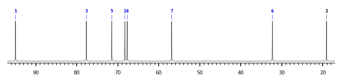
The spectrum also has 1 signal at unknown position (not plotted). |
There is only one chemically distinct structure:
OC(C)=O](smiles2image.php?&smiles=%2A%5B1%2A%5D%28%2A%29OC%28C%29%3DO&minwidth=240) R1 = bDSugp, Sug = 6-deoxy-Hepp
R1 = bDSugp, Sug = 6-deoxy-Hepp
*[1*](*)OC(C)=O
59.044 g/mol (C2H3R3O2, R = next and previous repeats / underdetermined substituent(s), not counted in mol weight)
 Found
Found  report error