Taxonomic group: bacteria / Proteobacteria
(Phylum: Proteobacteria)
Associated disease: infection due to Escherichia coli [ICD11:
XN6P4 
]
The structure was elucidated in this paperPublication DOI: 10.1016/S0008-6215(00)90931-5Journal NLM ID: 0043535Publisher: Elsevier
Institutions: School of Pharmaceutical Sciences, Rhodes University, Grahamstown 6140 South Africa
The capsular polysaccharide from Escherichia coli K47 was investigated using mainly methylation analysis and 1H and 13C NMR spectroscopy and shown to have the following repeating unit: (structure: see text).
Structure type: polymer chemical repeating unit
Location inside paper: p.199, structure 1
Compound class: CPS, O-polysaccharide, K-antigen, O-antigen
Contained glycoepitopes: IEDB_134623,IEDB_135813,IEDB_136044,IEDB_136906,IEDB_137340,IEDB_137472,IEDB_137485,IEDB_141794,IEDB_141807,IEDB_144983,IEDB_151528,IEDB_151531,IEDB_151771,IEDB_152206,IEDB_190606,IEDB_983930,SB_165,SB_166,SB_187,SB_195,SB_44,SB_7,SB_72,SB_88
Methods: 13C NMR, 1H NMR, GLC-MS, NMR-2D
Comments, role: published polymerization frame was shifted for conformity with other records.
NCBI Taxonomy refs (TaxIDs): 562
Show glycosyltransferases
NMR conditions: in D2O at 303 K
[as TSV]
13C NMR data:
Linkage	Residue	C1	C2	C3	C4	C5	C6
3,4,4,4	xRPyr
3,4,4	bDGalp	101.65	79.16	79.96	76.21	73.76	?
3,4	bDManp	101.65	70.83	72.39	78.25	75.71	?
3	aDGalp	100.11	69.30	70.10	76.97	71.18	60.88
2	Ac
	bDGlcpN	100.60	55.21	80.49	71.46	76.45	61.28


1H NMR data:
Linkage	Residue	H1	H2	H3	H4	H5	H6
3,4,4,4	xRPyr
3,4,4	bDGalp	4.62	3.81	4.39	4.29	4.07	3.84-3.91
3,4	bDManp	4.85	4.22	3.79	3.77	3.52	?
3	aDGalp	5.46	3.89	3.94	4.26	3.91	3.70-3.78
2	Ac
	bDGlcpN	4.91	3.86	3.77	3.74	3.45	3.76-3.76


1H/13C HSQC data:
Linkage	Residue	C1/H1	C2/H2	C3/H3	C4/H4	C5/H5	C6/H6
3,4,4,4	xRPyr
3,4,4	bDGalp	101.65/4.62	79.16/3.81	79.96/4.39	76.21/4.29	73.76/4.07	?/3.84-3.91
3,4	bDManp	101.65/4.85	70.83/4.22	72.39/3.79	78.25/3.77	75.71/3.52	?/?
3	aDGalp	100.11/5.46	69.30/3.89	70.10/3.94	76.97/4.26	71.18/3.91	60.88/3.70-3.78
2	Ac
	bDGlcpN	100.60/4.91	55.21/3.86	80.49/3.77	71.46/3.74	76.45/3.45	61.28/3.76-3.76
1H NMR data:
| Linkage | Residue | H1 | H2 | H3 | H4 | H5 | H6 |
|---|
| 3,4,4,4 | xRPyr | |
| 3,4,4 | bDGalp | 4.62 | 3.81 | 4.39 | 4.29 | 4.07 | 3.84
3.91 |
| 3,4 | bDManp | 4.85 | 4.22 | 3.79 | 3.77 | 3.52 | ? |
| 3 | aDGalp | 5.46 | 3.89 | 3.94 | 4.26 | 3.91 | 3.70
3.78 |
| 2 | Ac | |
| | bDGlcpN | 4.91 | 3.86 | 3.77 | 3.74 | 3.45 | 3.76
3.76 |
|
13C NMR data:
| Linkage | Residue | C1 | C2 | C3 | C4 | C5 | C6 |
|---|
| 3,4,4,4 | xRPyr | |
| 3,4,4 | bDGalp | 101.65 | 79.16 | 79.96 | 76.21 | 73.76 | ? |
| 3,4 | bDManp | 101.65 | 70.83 | 72.39 | 78.25 | 75.71 | ? |
| 3 | aDGalp | 100.11 | 69.30 | 70.10 | 76.97 | 71.18 | 60.88 |
| 2 | Ac | |
| | bDGlcpN | 100.60 | 55.21 | 80.49 | 71.46 | 76.45 | 61.28 |
|
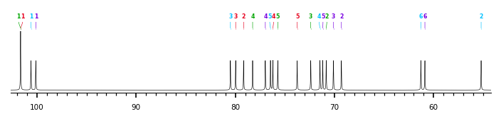
The spectrum also has 2 signals at unknown positions (not plotted). |
There is only one chemically distinct structure:
 Found
Found  report error