Taxonomic group: bacteria / Bacteroidetes
(Phylum: Bacteroidetes)
Host organism: Homo sapiens
Associated disease: periodontitis [ICD11:
DA0C 
]
The structure was elucidated in this paperNCBI PubMed ID: 27986835Publication DOI: 10.1093/glycob/cww129Journal NLM ID: 9104124Publisher: IRL Press at Oxford University Press
Correspondence: christina.schaeffer

boku.ac.at; Ian.Schoenhofen
nrc-cnrc.gc.ca
Institutions: Department of Oral Biology, School of Dental Medicine, University at Buffalo, 311 Foster Hall, 3435 Main St. Buffalo, New York 14214, USA, Department of Chemistry, Universitat fur Bodenkultur Wien, Muthgasse 18, A-1190 Vienna, Austria, National Research Council, Human Health Therapeutics Portfolio, 100 Sussex Drive, Ottawa, ON, Canada K1A 0R6, Department of NanoBiotechnology, NanoGlycobiology Unit, Universitat fur Bodenkultur Wien, Muthgasse 11, A-1190 Vienna, Austria
Tannerella forsythia is an anaerobic, Gram-negative periodontal pathogen. A unique O-linked oligosaccharide decorates the bacterium's cell surface proteins and was shown to modulate the host immune response. In our study, we investigated the biosynthesis of the nonulosonic acid (NulO) present at the terminal position of this glycan. A bioinformatic analysis of T. forsythia genomes revealed a gene locus for the synthesis of pseudaminic acid (Pse) in the type strain ATCC 43037 while strains FDC 92A2 and UB4 possess a locus for the synthesis of legionaminic acid (Leg) instead. In contrast to the NulO in ATCC 43037, which has been previously identified as a Pse derivative (5-N-acetimidoyl-7-N-glyceroyl-3,5,7,9-tetradeoxy-l-glycero-l-manno-NulO), glycan analysis of strain UB4 performed in this study indicated a 350-Da, possibly N-glycolyl Leg (3,5,7,9-tetradeoxy-d-glycero-d-galacto-NulO) derivative with unknown C5,7 N-acyl moieties. We have expressed, purified and characterized enzymes of both NulO pathways to confirm these genes' functions. Using capillary electrophoresis (CE), CE-mass spectrometry and NMR spectroscopy, our studies revealed that Pse biosynthesis in ATCC 43037 essentially follows the UDP-sugar route described in Helicobacter pylori, while the pathway in strain FDC 92A2 corresponds to Leg biosynthesis in Campylobacter jejuni involving GDP-sugar intermediates. To demonstrate that the NulO biosynthesis enzymes are functional in vivo, we created knockout mutants resulting in glycans lacking the respective NulO. Compared to the wild-type strains, the mutants exhibited significantly reduced biofilm formation on mucin-coated surfaces, suggestive of their involvement in host-pathogen interactions or host survival. This study contributes to understanding possible biological roles of bacterial NulOs.
Campylobacter jejuni, Helicobacter, Biofilm, biosynthesis pathway, bacterium, pseudaminic and legionaminic acid
Structure type: monomer
Location inside paper: p.345, fig.2, compound IIL, fig.6A, table II, GDP-2-acetamido-2,6-dideoxy-α-D-xylo-hexos-4-ulose
Trivial name: GDP-2-acetamido-2,6-α-D-xylo-hexos-4-ulose, GDP-2-acetamido-2,6-dideoxy-α-D-xylo-hexos-4-ulose
Contained glycoepitopes: IEDB_141493,IEDB_149170,IEDB_190357
Methods: 13C NMR, 1H NMR, PCR, SDS-PAGE, DNA techniques, b-elimination, genetic methods, biochemical methods, CE-MS, bioinformatic analysis, CE, LC-ESI-MS/MS, biofilm assays
Enzymes that release or process the structure: LegB (dehydratase BFO_1074-His6)
Synthetic data: enzymatic
Comments, role: The NMR temperature was not indicated
Related record ID(s): 11895, 12084, 12086, 12087, 12088
NCBI Taxonomy refs (TaxIDs): 203275
Show glycosyltransferases
NMR conditions: in D2O
[as TSV]
13C NMR data:
Linkage	Residue	C1	C2	C3	C4	C5	C6
5,0,0,2	Ac
5,0,0	aD6dxylHexpN-4-ulo	95.5	53.7	72.5	?	71.1	12.5
5,0	P
5	P
	xXnucG


1H NMR data:
Linkage	Residue	H1	H2	H3	H4	H5	H6
5,0,0,2	Ac
5,0,0	aD6dxylHexpN-4-ulo	5.46	4.09	3.83	-	4.11	1.21
5,0	P
5	P
	xXnucG


1H/13C HSQC data:
Linkage	Residue	C1/H1	C2/H2	C3/H3	C4/H4	C5/H5	C6/H6
5,0,0,2	Ac
5,0,0	aD6dxylHexpN-4-ulo	95.5/5.46	53.7/4.09	72.5/3.83		71.1/4.11	12.5/1.21
5,0	P
5	P
	xXnucG
1H NMR data:
| Linkage | Residue | H1 | H2 | H3 | H4 | H5 | H6 |
|---|
| 5,0,0,2 | Ac | |
| 5,0,0 | aD6dxylHexpN-4-ulo | 5.46 | 4.09 | 3.83 |
| 4.11 | 1.21 |
| 5,0 | P | |
| 5 | P | |
| | xXnucG | |
|
13C NMR data:
| Linkage | Residue | C1 | C2 | C3 | C4 | C5 | C6 |
|---|
| 5,0,0,2 | Ac | |
| 5,0,0 | aD6dxylHexpN-4-ulo | 95.5 | 53.7 | 72.5 | ? | 71.1 | 12.5 |
| 5,0 | P | |
| 5 | P | |
| | xXnucG | |
|
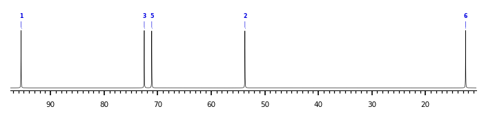
The spectrum also has 1 signal at unknown position (not plotted). |
There is only one chemically distinct structure:
 Found
Found  report error