Taxonomic group: bacteria / Firmicutes
(Phylum: Firmicutes)
Associated disease: infection due to Streptococcus pneumoniae [ICD11:
XN3PW 
]
The structure was elucidated in this paperNCBI PubMed ID: 9116006Publication DOI: 10.1021/bi961819lJournal NLM ID: 0370623Publisher: American Chemical Society
Institutions: Institute for Biological Sciences, National Research Council of Canada, Ottawa, Canada K1A 0R6, and Channing Laboratory, HarVard Medical School, Boston, Massachusetts 02115
The conformational epitope of the type III group B Streptococcus capsular polysaccharide (GBSP III) exhibits unique properties which can be ascribed to the presence of sialic acid in its structure and the requirement for an extended binding site. By means of NMR and molecular dynamics studies on GBSP III and its fragments, the extended epitope of GBSP III was further defined. The influence of sialic acid on the conformational properties of GBSP III was examined by performing conformational analysis on desialylated GBSP III, which is identical to the polysaccharide of Streptococcus pneumoniae type 14, and also on oxidized and reduced GBSP III. Conformational changes were gauged by 1H and 13C chemical shift analysis, NOE, 1D selective TOCSY-NOESY experiments, J(HH) and J(CH) variations, and NOE of OH resonances. Changes in mobility were examined by 13C T1 and T2 measurements. Unrestrained molecular dynamics simulations with explicit water using the AMBER force field and the GLYCAM parameter set were used to assess static and dynamic conformational models, simulate the observable NMR parameters and calculate helical parameters. GBSP III was found to be capable of forming extended helices. Hence, the length dependence of the conformational epitope could be explained by its location on extended helices within the random coil structure of GBSP III. The interaction of sialic acid with the backbone of the PS was also found to be important in defining the conformational epitope of GBSP III
NMR, capsular, polysaccharide, Streptococcus, group, molecular, epitope, capsular polysaccharide, type, conformational, dynamics, group B Streptococcus, molecular dynamics, NMR spectroscopy, type III group B streptococcus
Structure type: polymer chemical repeating unit
Location inside paper: Fig. 1, [c(d)ba]n
Compound class: CPS, O-polysaccharide, O-antigen
Contained glycoepitopes: IEDB_130646,IEDB_130697,IEDB_135813,IEDB_136044,IEDB_137340,IEDB_137472,IEDB_137776,IEDB_1391966,IEDB_1392542,IEDB_140108,IEDB_140110,IEDB_140122,IEDB_141794,IEDB_141807,IEDB_142351,IEDB_142487,IEDB_142488,IEDB_143634,IEDB_146664,IEDB_149138,IEDB_149139,IEDB_149141,IEDB_149142,IEDB_149143,IEDB_149144,IEDB_149145,IEDB_149147,IEDB_149148,IEDB_149149,IEDB_149150,IEDB_149151,IEDB_151531,IEDB_190606,IEDB_983931,SB_145,SB_165,SB_166,SB_173,SB_187,SB_192,SB_195,SB_30,SB_6,SB_7,SB_88
Methods: 13C NMR, 1H NMR, NMR-2D, enzymatic hydrolysis
Comments, role: [c(d)ba]n is desialylated capsular polysaccharide, the repeating unit has the same structure as Streptococcus pneumonia type 14 capsular polysaccharide
3D data: molecular dynamics, molecular modeling
Related record ID(s): 1658, 1798, 1799, 1800, 1801, 1802, 1803
NCBI Taxonomy refs (TaxIDs): 1313Reference(s) to other database(s): GTC:G52755VD, GlycomeDB:
2039, CCSD:
11172, CBank-STR:12723
Show glycosyltransferases
NMR conditions: in D2O at 290 K
[as TSV]
13C NMR data:
Linkage	Residue	C1	C2	C3	C4	C5	C6
3,6	bDGlcp	103.44	73.47	75.08	79.14	75.56	60.85
3,2	Ac	?	?
3,4	bDGalp	103.52	71.74	73.28	69.42	76.10	61.95
3	bDGlcpN	103.81	56.01	72.84	78.21	74.07	68.25
	bDGalp	103.71	70.80	83.06	69.07	75.76	61.84


1H NMR data:
Linkage	Residue	H1	H2	H3	H4	H5	H6
3,6	bDGlcp	4.55	3.37	3.65	3.64	3.61	3.79-3.98
3,2	Ac
3,4	bDGalp	4.55	3.54	3.67	3.92	3.69	3.75-3.77
3	bDGlcpN	4.70	3.81	3.71	3.91	3.71	3.98-4.27
	bDGalp	4.43	3.58	3.73	4.17	3.69	3.75-3.78


1H/13C HSQC data:
Linkage	Residue	C1/H1	C2/H2	C3/H3	C4/H4	C5/H5	C6/H6
3,6	bDGlcp	103.44/4.55	73.47/3.37	75.08/3.65	79.14/3.64	75.56/3.61	60.85/3.79-3.98
3,2	Ac	NMR TSV error 2: unequal length of 13C and 1H datasets
3,4	bDGalp	103.52/4.55	71.74/3.54	73.28/3.67	69.42/3.92	76.10/3.69	61.95/3.75-3.77
3	bDGlcpN	103.81/4.70	56.01/3.81	72.84/3.71	78.21/3.91	74.07/3.71	68.25/3.98-4.27
	bDGalp	103.71/4.43	70.80/3.58	83.06/3.73	69.07/4.17	75.76/3.69	61.84/3.75-3.78
1H NMR data:
| Linkage | Residue | H1 | H2 | H3 | H4 | H5 | H6 |
|---|
| 3,6 | bDGlcp | 4.55 | 3.37 | 3.65 | 3.64 | 3.61 | 3.79
3.98 |
| 3,2 | Ac | |
| 3,4 | bDGalp | 4.55 | 3.54 | 3.67 | 3.92 | 3.69 | 3.75
3.77 |
| 3 | bDGlcpN | 4.70 | 3.81 | 3.71 | 3.91 | 3.71 | 3.98
4.27 |
| | bDGalp | 4.43 | 3.58 | 3.73 | 4.17 | 3.69 | 3.75
3.78 |
|
13C NMR data:
| Linkage | Residue | C1 | C2 | C3 | C4 | C5 | C6 |
|---|
| 3,6 | bDGlcp | 103.44 | 73.47 | 75.08 | 79.14 | 75.56 | 60.85 |
| 3,2 | Ac | ? | ? | |
| 3,4 | bDGalp | 103.52 | 71.74 | 73.28 | 69.42 | 76.10 | 61.95 |
| 3 | bDGlcpN | 103.81 | 56.01 | 72.84 | 78.21 | 74.07 | 68.25 |
| | bDGalp | 103.71 | 70.80 | 83.06 | 69.07 | 75.76 | 61.84 |
|
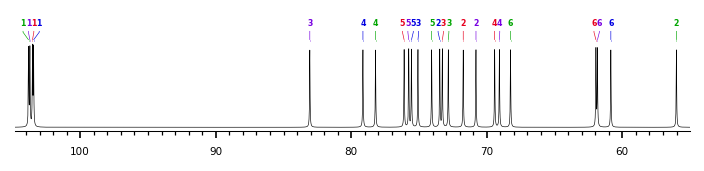
The spectrum also has 2 signals at unknown positions (not plotted). |
There is only one chemically distinct structure:
 report error
 Found 1 record.
Displayed record 1
Found 1 record.
Displayed record 1
