Taxonomic group: bacteria / Proteobacteria
(Phylum: Proteobacteria)
Associated disease: infection due to Escherichia coli [ICD11:
XN6P4 
]
The structure was elucidated in this paperNCBI PubMed ID: 9652410Journal NLM ID: 0107600Publisher: Oxford, UK: Blackwell Science Ltd. on behalf of the Federation of European Biochemical Societies
Correspondence: gw

admin.organ.su.se
Institutions: Department of Organic Chemistry, Arrhenius Laboratory, Stockholm University, Sweden
The structure of the O-antigenic polysaccharide from Escherichia coli O141 has been determined. NMR spectroscopy and sugar and methylation analyses were the principal methods used. The sequence of the sugar residues could be determined by NOESY and heteronuclear multiple-bond connectivity (HMBC-) NMR experiments. The polysaccharide is composed of pentasaccharide repeating units with 1 O-acetyl group/repeating unit. The following structure, where Rha is 6-deoxymannose is concluded: carbohydrate sequence [see text]
Lipopolysaccharide, NMR, structure, O-antigen, enterotoxigenic Escherichia coli
Structure type: suggested polymer biological repeating unit
Location inside paper: abstract
Compound class: O-polysaccharide, O-antigen
Contained glycoepitopes: IEDB_115136,IEDB_130701,IEDB_135813,IEDB_136105,IEDB_137340,IEDB_140630,IEDB_141807,IEDB_144983,IEDB_151531,IEDB_152206,IEDB_164174,IEDB_225177,IEDB_423153,IEDB_885823,IEDB_983930,SB_197,SB_44,SB_67,SB_72
Methods: methylation, NMR-2D, NMR, sugar analysis, de-O-acetylation
Comments, role: chemical repeat frame is different in the paper
Related record ID(s): 20684
NCBI Taxonomy refs (TaxIDs): 562Reference(s) to other database(s): GTC:G84496XY, GlycomeDB:
28125
Show glycosyltransferases
NMR conditions: in D2O at 303 K
[as TSV]
13C NMR data:
Linkage	Residue	C1	C2	C3	C4	C5	C6
3,3,2	bDGlcpA	104.1	73.8	76.7	73.0	77.3	?
3,3,3	aLRhap	98.0	71.8	71.6	73.7	70.3	17.4
3,3	aDManp	101.5	75.6	72.8	72.9	74.2	62.3
3,6	Ac	?	21.0
3	aDManp	102.8	71.4	80.1	67.0	72.6	64.8
2	Ac	?	23.1
	bDGlcpN	102.2	56.2	82.1	72.6	77.6	62.9


1H NMR data:
Linkage	Residue	H1	H2	H3	H4	H5	H6
3,3,2	bDGlcpA	4.52	3.38	3.51	3.62	3.86	-
3,3,3	aLRhap	4.94	3.98	3.95	3.47	4.25	1.31
3,3	aDManp	5.20	4.37	4.09	3.95	3.82	3.82
3,6	Ac	-	2.16
3	aDManp	5.18	4.15	3.90	3.87	3.76	4.28-4.40
2	Ac	-	2.04
	bDGlcpN	5.45	3.79	3.69	3.49	3.44	3.75-3.94


1H/13C HSQC data:
Linkage	Residue	C1/H1	C2/H2	C3/H3	C4/H4	C5/H5	C6/H6
3,3,2	bDGlcpA	104.1/4.52	73.8/3.38	76.7/3.51	73.0/3.62	77.3/3.86
3,3,3	aLRhap	98.0/4.94	71.8/3.98	71.6/3.95	73.7/3.47	70.3/4.25	17.4/1.31
3,3	aDManp	101.5/5.20	75.6/4.37	72.8/4.09	72.9/3.95	74.2/3.82	62.3/3.82
3,6	Ac		21.0/2.16
3	aDManp	102.8/5.18	71.4/4.15	80.1/3.90	67.0/3.87	72.6/3.76	64.8/4.28-4.40
2	Ac		23.1/2.04
	bDGlcpN	102.2/5.45	56.2/3.79	82.1/3.69	72.6/3.49	77.6/3.44	62.9/3.75-3.94
1H NMR data:
| Linkage | Residue | H1 | H2 | H3 | H4 | H5 | H6 |
|---|
| 3,3,2 | bDGlcpA | 4.52 | 3.38 | 3.51 | 3.62 | 3.86 |
|
| 3,3,3 | aLRhap | 4.94 | 3.98 | 3.95 | 3.47 | 4.25 | 1.31 |
| 3,3 | aDManp | 5.20 | 4.37 | 4.09 | 3.95 | 3.82 | 3.82 |
| 3,6 | Ac |
| 2.16 | |
| 3 | aDManp | 5.18 | 4.15 | 3.90 | 3.87 | 3.76 | 4.28
4.40 |
| 2 | Ac |
| 2.04 | |
| | bDGlcpN | 5.45 | 3.79 | 3.69 | 3.49 | 3.44 | 3.75
3.94 |
|
13C NMR data:
| Linkage | Residue | C1 | C2 | C3 | C4 | C5 | C6 |
|---|
| 3,3,2 | bDGlcpA | 104.1 | 73.8 | 76.7 | 73.0 | 77.3 | ? |
| 3,3,3 | aLRhap | 98.0 | 71.8 | 71.6 | 73.7 | 70.3 | 17.4 |
| 3,3 | aDManp | 101.5 | 75.6 | 72.8 | 72.9 | 74.2 | 62.3 |
| 3,6 | Ac | ? | 21.0 | |
| 3 | aDManp | 102.8 | 71.4 | 80.1 | 67.0 | 72.6 | 64.8 |
| 2 | Ac | ? | 23.1 | |
| | bDGlcpN | 102.2 | 56.2 | 82.1 | 72.6 | 77.6 | 62.9 |
|
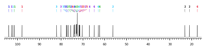
The spectrum also has 3 signals at unknown positions (not plotted). |
There is only one chemically distinct structure:
 report error
 Found 1 record.
Displayed record 1
Found 1 record.
Displayed record 1
