Taxonomic group: bacteria / Proteobacteria
(Phylum: Proteobacteria)
Associated disease: infection due to Escherichia coli [ICD11:
XN6P4 
]
The structure was elucidated in this paperPublication DOI: 10.1080/07328300903296394Journal NLM ID: 8218151Publisher: Marcel Dekker
Correspondence: perepel

ioc.ac.ru
Institutions: N.D. Zelinsky Institute of Organic Chemistry, Russian Academy of Sciences, Moscow, Russia
On mild acid degradation of the lipopolysaccharide of Escherichia coli O61, the O-polysaccharide chain was cleaved at a linkage of 5,7-diacetamido-3,5,7,9-tetradeoxy-l-glycero-D-galacto-non-2-ulosonic acid (di-N-acetyl-8-epilegionaminic acid, 8eLeg5Ac7Ac). The resultant trisaccharide, an O-deacylated lipopolysaccharide and an O-deacetylated trisaccharide derived from the latter were studied by sugar analyses along with 1H and 13C NMR spectroscopy, and the following structure of the O-polysaccharide was established: [formula, see text].
Lipopolysaccharide, O-antigen, Escherichia coli, nonulosonic acid, bacterial polysaccharide structure, 8-epilegionaminic acid
Structure type: polymer chemical repeating unit
Location inside paper: p.464, p.470
Compound class: O-polysaccharide, O-antigen
Contained glycoepitopes: IEDB_130648,IEDB_137473,IEDB_1391961,IEDB_141584,IEDB_142488,IEDB_146664,IEDB_885822,IEDB_983931,SB_192
Methods: 13C NMR, 1H NMR, NMR-2D, sugar analysis, GLC, alkaline degradation, mild acid degradation
Comments, role: The signals for NAc are 1.95-2.03; 23.3-23.7(Me) and 175.0-175.9(CO); 1.98-2.02, 23.3-23.7 (Me) and 175.1-176.0 (CO); 1.99 (Me); 23.0-23.6 (Me) and 175.1-176.0 (CO).
NCBI Taxonomy refs (TaxIDs): 562Reference(s) to other database(s): GTC:G67059IO
Show glycosyltransferases
NMR conditions: in D2O at 303 K
[as TSV]
13C NMR data:
Linkage	Residue	C1	C2	C3	C4	C5	C6	C7	C8	C9
3,4,5	Ac
3,4,7	Ac
3,4,8	70%Ac	?	21.9
3,4	aX8eLegp	?	101.9	37.6	72.4	51.7	75.2	55.0	69.5	20.2
3	bDGlcp	105.8	74.3	76.2	73.2	76.2	61.3
2	Ac
	aDGalpN	94.6	49.4	79.2	70.0	73.2	62.2


1H NMR data:
Linkage	Residue	H1	H2	H3	H4	H5	H6	H7	H8	H9
3,4,5	Ac
3,4,7	Ac
3,4,8	70%Ac	-	2.00
3,4	aX8eLegp	-	-	1.63-2.78	3.72	3.72	3.86	3.91	3.98	1.22
3	bDGlcp	4.45	3.27	3.51	3.98	3.41	3.76-3.84
2	Ac
	aDGalpN	5.01	3.34	3.70	4.17	3.72	3.69-3.71


1H/13C HSQC data:
Linkage	Residue	C1/H1	C2/H2	C3/H3	C4/H4	C5/H5	C6/H6	C7/H7	C8/H8	C9/H9
3,4,5	Ac
3,4,7	Ac
3,4,8	70%Ac		21.9/2.00
3,4	aX8eLegp			37.6/1.63-2.78	72.4/3.72	51.7/3.72	75.2/3.86	55.0/3.91	69.5/3.98	20.2/1.22
3	bDGlcp	105.8/4.45	74.3/3.27	76.2/3.51	73.2/3.98	76.2/3.41	61.3/3.76-3.84
2	Ac
	aDGalpN	94.6/5.01	49.4/3.34	79.2/3.70	70.0/4.17	73.2/3.72	62.2/3.69-3.71
1H NMR data:
| Linkage | Residue | H1 | H2 | H3 | H4 | H5 | H6 | H7 | H8 | H9 |
|---|
| 3,4,5 | Ac | |
| 3,4,7 | Ac | |
| 3,4,8 | 70%Ac |
| 2.00 | |
| 3,4 | aX8eLegp |
|
| 1.63
2.78 | 3.72 | 3.72 | 3.86 | 3.91 | 3.98 | 1.22 |
| 3 | bDGlcp | 4.45 | 3.27 | 3.51 | 3.98 | 3.41 | 3.76
3.84 | |
| 2 | Ac | |
| | aDGalpN | 5.01 | 3.34 | 3.70 | 4.17 | 3.72 | 3.69
3.71 | |
|
13C NMR data:
| Linkage | Residue | C1 | C2 | C3 | C4 | C5 | C6 | C7 | C8 | C9 |
|---|
| 3,4,5 | Ac | |
| 3,4,7 | Ac | |
| 3,4,8 | 70%Ac | ? | 21.9 | |
| 3,4 | aX8eLegp | ? | 101.9 | 37.6 | 72.4 | 51.7 | 75.2 | 55.0 | 69.5 | 20.2 |
| 3 | bDGlcp | 105.8 | 74.3 | 76.2 | 73.2 | 76.2 | 61.3 | |
| 2 | Ac | |
| | aDGalpN | 94.6 | 49.4 | 79.2 | 70.0 | 73.2 | 62.2 | |
|
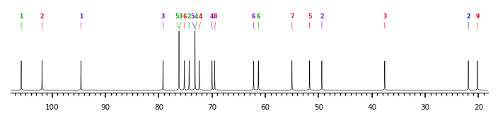
The spectrum also has 2 signals at unknown positions (not plotted). |
There is only one chemically distinct structure:
 Found
Found  report error