Taxonomic group: bacteria / Proteobacteria
(Phylum: Proteobacteria)
Associated disease: cystic fibrosis (CF) [ICD11:
CA25 
];
infection due to Pseudomonas aeruginosa [ICD11:
XN5L6 ]
The structure was elucidated in this paperNCBI PubMed ID: 21151973Journal NLM ID: 101285081Publisher: San Francisco, CA: Public Library of Science
Correspondence: Irina.Sadovskaya

univ-littoral.fr
Institutions: Universite Lille Nord de France, Lille, France
Background: Pseudomonas aeruginosa is a Gram-negative bacterium and an opportunistic pathogen, which causespersisting life-threatening infections in cystic fibrosis (CF) patients. Biofilm mode of growth facilitates its survival in a variety of environments. Most P. aeruginosa isolates, including the non-mucoid laboratory strain PA14, are able to form a thick pellicle, which results in a surface-associated biofilm at the air-liquid (A-L) interface in standing liquid cultures.Exopolysaccharides (EPS) are considered as key components in the formation of this biofilm pellicle. In the non-mucoid P.aeruginosa strain PA14, the "scaffolding" polysaccharides of the biofilm matrix, and the molecules responsible for the structural integrity of rigid A-L biofilm have not been identified. Moreover, the role of LPS in this process is unclear, and the chemical structure of the LPS O-antigen of PA14 has not yet been elucidated. Principal Findings: In the present work we carried out a systematic analysis of cellular and extracellular (EC) carbohydrates of P. aeruginosa PA14. We also elucidated the chemical structure of the LPS O-antigen by chemical methods and 2-D NMR spectroscopy. Our results showed that it is composed of linear trisaccharide repeating units, identical to those described for P. aeruginosa Lanyi type O:2a,c (Lanyi-Bergman O-serogroup 10a, 10c; IATS serotype 19) and having the following structure:-4)-a-L-GalNAcA-(1-3)-a-D-QuiNAc-(1-3)-a-L-Rha-(1-. Furthermore, an EC O-antigen polysaccharide (EC O-PS) and the glycerol-phosphorylated cyclic b-(1,3)-glucans were identified in the culture supernatant of PA14, grown statically inminimal medium. Finally, the extracellular matrix of the thick biofilm formed at the A-L interface contained, in addition to eDNA, important quantities (at least, 20% of dry weight) of LPS-like material. Conclusions: We characterized the chemical structure of the LPS O-antigen and showed that the O-antigen polysaccharide is an abundant extracellular carbohydrate of PA14. We present evidence that LPS-like material is found as a component of a biofilm matrix of P. aeruginosa.
Pseudomonas aeruginosa, NMR spectroscopy, exopolysaccharides, cystic fibrosis, Biofilm
Structure type: polymer chemical repeating unit
Location inside paper: p.3, fig.1B
Compound class: EPS, O-polysaccharide, O-antigen
Contained glycoepitopes: IEDB_130667,IEDB_136105,IEDB_225177,IEDB_885823
Methods: 13C NMR, 1H NMR, methylation, NMR-2D, GC-MS, DOC-PAGE, GC, composition analysis, NMR-1D, HF treatment
Comments, role: life-threatening infections in cystic fibrosis (CF) patients; published polymerization frame was shifted for conformity with other records.
Related record ID(s): 108468, 122632, 143843
NCBI Taxonomy refs (TaxIDs): 652611Reference(s) to other database(s): GTC:G07327IC, GlycomeDB:
3411
Show glycosyltransferases
NMR conditions: in D2O at 298 K
[as TSV]
13C NMR data:
Linkage	Residue	C1	C2	C3	C4	C5	C6
3,4	aLRhap	102.3	68.8	77.1	71.4	70.5	17.8
3,2	Ac
3	aLGalpNA	98.2	50.7	67.6	80.0	71.6	?
2	Ac
	aDQuipN	95.9	54.9	76.4	75.2	69.2	17.8


1H NMR data:
Linkage	Residue	H1	H2	H3	H4	H5	H6
3,4	aLRhap	4.84	3.95	3.78	3.50	4.05	1.26
3,2	Ac
3	aLGalpNA	5.16	4.17	4.10	4.40	4.97	-
2	Ac
	aDQuipN	4.87	4.12	3.90	3.28	4.10	1.26


1H/13C HSQC data:
Linkage	Residue	C1/H1	C2/H2	C3/H3	C4/H4	C5/H5	C6/H6
3,4	aLRhap	102.3/4.84	68.8/3.95	77.1/3.78	71.4/3.50	70.5/4.05	17.8/1.26
3,2	Ac
3	aLGalpNA	98.2/5.16	50.7/4.17	67.6/4.10	80.0/4.40	71.6/4.97
2	Ac
	aDQuipN	95.9/4.87	54.9/4.12	76.4/3.90	75.2/3.28	69.2/4.10	17.8/1.26
1H NMR data:
| Linkage | Residue | H1 | H2 | H3 | H4 | H5 | H6 |
|---|
| 3,4 | aLRhap | 4.84 | 3.95 | 3.78 | 3.50 | 4.05 | 1.26 |
| 3,2 | Ac | |
| 3 | aLGalpNA | 5.16 | 4.17 | 4.10 | 4.40 | 4.97 |
|
| 2 | Ac | |
| | aDQuipN | 4.87 | 4.12 | 3.90 | 3.28 | 4.10 | 1.26 |
|
13C NMR data:
| Linkage | Residue | C1 | C2 | C3 | C4 | C5 | C6 |
|---|
| 3,4 | aLRhap | 102.3 | 68.8 | 77.1 | 71.4 | 70.5 | 17.8 |
| 3,2 | Ac | |
| 3 | aLGalpNA | 98.2 | 50.7 | 67.6 | 80.0 | 71.6 | ? |
| 2 | Ac | |
| | aDQuipN | 95.9 | 54.9 | 76.4 | 75.2 | 69.2 | 17.8 |
|
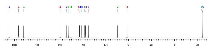
The spectrum also has 1 signal at unknown position (not plotted). |
There is only one chemically distinct structure:
 report error
 Found 1 record.
Displayed record 1
Found 1 record.
Displayed record 1
