Taxonomic group: bacteria / Firmicutes
(Phylum: Firmicutes)
Associated disease: infection due to Streptococcus pneumoniae [ICD11:
XN3PW 
]
The structure was elucidated in this paperNCBI PubMed ID: 11841814Journal NLM ID: 0043535Publisher: Elsevier
Correspondence: bush+AEA-umbc.edu
Institutions: Department of Chemistry and Biochemistry, University of Maryland Baltimore County, 1000 Hilltop Circle, Baltimore, MD 21250, USA, Bioprocess and Bioanalytical Chemistry, Merck Research Laboratories, West Point, PA 19486, USA
NMR spectroscopy can be used to characterize bacterial polysaccharides such as that of Streptococcus pneumoniae type 1 which is a component of the 23-valent pneumococcal vaccine in clinical use. This particular polysaccharide gives NMR spectra with wide lines apparently due to restricted molecular mobility and chain flexibility which leads to rapid dipolar T(2) relaxation limiting the possibility of detailed spectral analysis. Removal of O-acetyl groups found on approximately two thirds of the repeating subunits of pneumococcal type 1 capsule leads to narrower NMR lines facilitating a complete assignment of the 1H and 13C NMR spectra. Degradation of the polysaccharide by periodate oxidation followed by base treatment leads to an oligosaccharide fragment of approximately three repeating trisaccharide units. This oligosaccharide has narrow NMR lines and 1H and 13C assignments very similar to those of the O-deacetylated polysaccharide. In the native polysaccharide, O-acetyl groups are located on the 2- and 3-positions of the 4-linked galacturonic acid residue providing protection against periodate oxidation. Analysis of NOESY spectra combined with molecular modeling of the oligosaccharide shows that flexibility occurs in certain of the saccharide linkages.
NMR, chemistry, structural, capsular, polysaccharide, Streptococcus, Streptococcus pneumoniae, analysis, capsular polysaccharide, type, structural analysis, chemical, biochemistry, vaccine, depolymerization
Structure type: polymer chemical repeating unit
Location inside paper: p.335
Compound class: CPS
Contained glycoepitopes: IEDB_142345
Methods: 13C NMR, 1H NMR, periodate oxidation, de-O-acetylation
Comments, role: NMR data of O-deacetylated polysaccharide
3D data: conformation data, molecular modeling
NCBI Taxonomy refs (TaxIDs): 1313Reference(s) to other database(s): GTC:G36906IX, GlycomeDB:
26501
Show glycosyltransferases
NMR conditions: in D2O at 303 K
[as TSV]
13C NMR data:
Linkage	Residue	C1	C2	C3	C4	C5	C6
3,3,2	Ac
3,3,3	Ac
3,3	aDGalpA	97.2	68.6	69.5	79.9	71.8	?
3	aDGalpA	99.5	66.6	76.6	68.5	72.8	?
2	Ac	?	22.8
	aDFucpN4N	99.1	48.4	73.8	53.6	63.5	15.8


1H NMR data:
Linkage	Residue	H1	H2	H3	H4	H5	H6
3,3,2	Ac
3,3,3	Ac
3,3	aDGalpA	5.22	3.92	4.07	4.38	4.58	-
3	aDGalpA	5.07	3.96	3.99	4.47	4.12	-
2	Ac	-	2.03
	aDFucpN4N	4.98	4.14	4.26	3.80	4.74	1.27


1H/13C HSQC data:
Linkage	Residue	C1/H1	C2/H2	C3/H3	C4/H4	C5/H5	C6/H6
3,3,2	Ac
3,3,3	Ac
3,3	aDGalpA	97.2/5.22	68.6/3.92	69.5/4.07	79.9/4.38	71.8/4.58
3	aDGalpA	99.5/5.07	66.6/3.96	76.6/3.99	68.5/4.47	72.8/4.12
2	Ac		22.8/2.03
	aDFucpN4N	99.1/4.98	48.4/4.14	73.8/4.26	53.6/3.80	63.5/4.74	15.8/1.27
1H NMR data:
| Linkage | Residue | H1 | H2 | H3 | H4 | H5 | H6 |
|---|
| 3,3,2 | Ac | |
| 3,3,3 | Ac | |
| 3,3 | aDGalpA | 5.22 | 3.92 | 4.07 | 4.38 | 4.58 |
|
| 3 | aDGalpA | 5.07 | 3.96 | 3.99 | 4.47 | 4.12 |
|
| 2 | Ac |
| 2.03 | |
| | aDFucpN4N | 4.98 | 4.14 | 4.26 | 3.80 | 4.74 | 1.27 |
|
13C NMR data:
| Linkage | Residue | C1 | C2 | C3 | C4 | C5 | C6 |
|---|
| 3,3,2 | Ac | |
| 3,3,3 | Ac | |
| 3,3 | aDGalpA | 97.2 | 68.6 | 69.5 | 79.9 | 71.8 | ? |
| 3 | aDGalpA | 99.5 | 66.6 | 76.6 | 68.5 | 72.8 | ? |
| 2 | Ac | ? | 22.8 | |
| | aDFucpN4N | 99.1 | 48.4 | 73.8 | 53.6 | 63.5 | 15.8 |
|
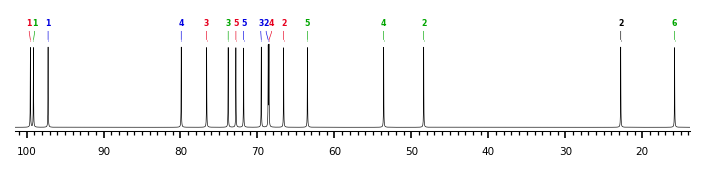
The spectrum also has 3 signals at unknown positions (not plotted). |
There is only one chemically distinct structure:
 report error
 Found 1 record.
Displayed record 1
Found 1 record.
Displayed record 1
