Taxonomic group: fungi / Mucoromycota
(Phylum: Mucoromycota)
NCBI PubMed ID: 30755333Publication DOI: 10.1016/j.bioorg.2019.01.046Journal NLM ID: 1303703Publisher: Orlando, FL: Academic Press
Correspondence: Sordon S <sandra.sordon

upwr.edu.pl>; Popłoński J <jaroslaw.poplonski
upwr.edu.pl>; Tronina T <tomasz.tronina
upwr.edu.pl>; Huszcza E <ewa.huszcza
upwr.edu.pl>
Institutions: Department of Chemistry, Wrocław University of Environmental and Life Sciences, Wrocław, Poland
In the present study, the species: Beauveria bassiana, Absidia coerulea and Absidia glauca were used in biotransformation of flavones (chrysin, apigenin, luteolin, diosmetin) and flavanones (pinocembrin, naringenin, eriodictyol, hesperetin). The Beauveria bassiana AM 278 strain catalyzed the methylglucose attachment reactions to the flavonoid molecule at positions C7 and C3'. The application of the Absidia genus (A. coerulea AM 93, A. glauca AM 177) as the biocatalyst resulted in the formation of glucosides with a sugar molecule present at C7 and C3' positions of flavonoids skeleton. Nine of obtained products have not been previously reported in the literature.
biotransformation, Beauveria bassiana, Absidia coerulea, microbial glycosylation, Absidia glauca, flavanones, flavones
Structure type: monomer
Location inside paper: Fig. 2, 12, supplementary materials, NMR data, 12
Trivial name: cosmosiin, apigenin 7-O-glucoside, rhoifolin, apigenin 7-glucoside, apigetrin
Compound class: glycoside, flavonoid glycoside, flavonol glycoside
Contained glycoepitopes: IEDB_142488,IEDB_146664,IEDB_613414,IEDB_983931,SB_192
Methods: 13C NMR, 1H NMR, NMR-2D, TLC, HPLC, biosynthetic methods, extraction, cell growth, HR-ESI-MS
Comments, role: NMR temperature was not specified
Related record ID(s): 49674, 49675, 49676, 49677, 49678, 49679, 49680, 49681, 49682, 49683, 49684, 49685, 49686, 49688, 49689, 49690, 49691, 49692
NCBI Taxonomy refs (TaxIDs): 90261,
4829Reference(s) to other database(s): CCSD:
50502, CBank-STR:1305
Show glycosyltransferases
NMR conditions: in DMSO-d6
[as TSV]
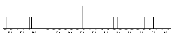
13C NMR data:
Linkage	Residue	C1	C2	C3	C4	C5	C6	C7	C8	C9	C10	C11	C12	C13	C14	C15	C16
7	bDGlcp	99.9	73.1	76.4	69.5	77.2	60.6
	xXApigenin	-	164.3	103.1	182.0	156.9	94.8	163.0	99.5	161.1	105.3	121.0	128.6	116.0	161.4	116.0	128.6


1H NMR data:
Linkage	Residue	H1	H2	H3	H4	H5	H6	H7	H8	H9	H10	H11	H12	H13	H14	H15	H16
7	bDGlcp	5.07	3.26	3.30	3.18	3.44	3.48-3.71
	xXApigenin	-	-	6.87	-	-	6.83	-	6.44	-	-	-	7.96	6.94	-	6.94	7.96


1H/13C HSQC data:
Linkage	Residue	C1/H1	C2/H2	C3/H3	C4/H4	C5/H5	C6/H6	C7/H7	C8/H8	C9/H9	C10/H10	C11/H11	C12/H12	C13/H13	C14/H14	C15/H15	C16/H16
7	bDGlcp	99.9/5.07	73.1/3.26	76.4/3.30	69.5/3.18	77.2/3.44	60.6/3.48-3.71
	xXApigenin			103.1/6.87			94.8/6.83		99.5/6.44				128.6/7.96	116.0/6.94		116.0/6.94	128.6/7.96
1H NMR data:
| Linkage | Residue | H1 | H2 | H3 | H4 | H5 | H6 | H7 | H8 | H9 | H10 | H11 | H12 | H13 | H14 | H15 | H16 |
|---|
| 7 | bDGlcp | 5.07 | 3.26 | 3.30 | 3.18 | 3.44 | 3.48
3.71 | |
| | xXApigenin |
|
| 6.87 |
|
| 6.83 |
| 6.44 |
|
|
| 7.96 | 6.94 |
| 6.94 | 7.96 |
|
13C NMR data:
| Linkage | Residue | C1 | C2 | C3 | C4 | C5 | C6 | C7 | C8 | C9 | C10 | C11 | C12 | C13 | C14 | C15 | C16 |
|---|
| 7 | bDGlcp | 99.9 | 73.1 | 76.4 | 69.5 | 77.2 | 60.6 | |
| | xXApigenin |
| 164.3 | 103.1 | 182.0 | 156.9 | 94.8 | 163.0 | 99.5 | 161.1 | 105.3 | 121.0 | 128.6 | 116.0 | 161.4 | 116.0 | 128.6 |
|
The spectrum also has 1 signal at unknown position (not plotted). |
There is only one chemically distinct structure:
 Found
Found  report error