Taxonomic group: bacteria / Proteobacteria
(Phylum: Proteobacteria)
Associated disease: infection due to Acinetobacter baumannii [ICD11:
XN8LS 
]
The structure was elucidated in this paperNCBI PubMed ID: 30664967Publication DOI: 10.1016/j.ijbiomac.2019.01.080Journal NLM ID: 7909578Publisher: Butterworth-Heinemann
Correspondence: J.J. Kenyon <johanna.kenyon

qut.edu.au>
Institutions: N.D. Zelinsky Institute of Organic Chemistry, Russian Academy of Sciences, Moscow, Russia, M. M. Shemyakin & Y. A. Ovchinnikov Institute of Bioorganic Chemistry, Russian Academy of Sciences, Moscow, Russia, State Research Center for Applied Microbiology and Biotechnology, Obolensk, Moscow Region, Russia, Moscow Institute of Physics and Technology, Dolgoprudny, Moscow Region, Russia, Institute of Health and Biomedical Innovation, School of Biomedical Sciences, Faculty of Health, Queensland University of Technology, Brisbane, Australia, School of Life and Environmental Sciences, The University of Sydney, Sydney, Australia, Institute of Antimicrobial Chemotherapy, Smolensk State Medical University, Smolensk, Russia
A new capsular polysaccharide (CPS) biosynthesis gene cluster, KL16, was found in the genome sequence of a clinical Acinetobacter baumannii ST25 isolate, D4. The variable part of KL16 contains a module of genes for synthesis of 5,7-diacetamido-3,5,7,9-tetradeoxy-l-glycero-l-manno-non-2-ulosonic acid (5,7-di-N-acetylpseudaminic acid, Pse5Ac7Ac), a gene encoding ItrA3 that initiates the CPS synthesis with d-GlcpNAc, and two glycosyltransferase (Gtr) genes. The K16 CPS was studied by sugar analysis and Smith degradation along with 1D and 2D 1H and 13C NMR spectroscopy, and shown to be built up of linear trisaccharide repeats containing d-galactose (d-Gal), N-acetyl-d-glucosamine (d-GlcNAc), and Pse5Ac7Ac. The d-Galp residue is linked to the d-GlcpNAc initiating sugar via a β-(1→3) linkage evidently formed by a Gtr5 variant, Gtr5K16, encoded in KL16. This reveals an altered or relaxed substrate specificity of this variant as the majority of Gtr5-type glycosyltransferases have previously been shown to form a β-d-Galp-(1→3)-d-GalpNAc linkage. The β-Psep5Ac7Ac-(2→4)-d-Galp linkage is predicted to be formed by the other glycosyltransferase, Gtr37, which does not match members of any known glycosyltransferase family.
Acinetobacter baumannii, capsular polysaccharide, 5, glycosyltransferase, 7-Di-N-acetylpseudaminic acid, KL16 K locus
Structure type: polymer chemical repeating unit
Location inside paper: table 1, p.104, fig.3, fig.4B, K16 CPS
Compound class: CPS
Contained glycoepitopes: IEDB_135813,IEDB_136044,IEDB_137340,IEDB_137472,IEDB_1391962,IEDB_141794,IEDB_141807,IEDB_142078,IEDB_143794,IEDB_150899,IEDB_151531,IEDB_190606,SB_137,SB_165,SB_166,SB_187,SB_195,SB_29,SB_7,SB_88
Methods: 13C NMR, 1H NMR, NMR-2D, sugar analysis, GLC, Smith degradation, function analysis of gene clusters
Enzymes that release or process the structure: Wzy(K16) polymerase, Gtr37, Gtr5(K16) glycosyltransferases, Wzy(K16)[ItrA3]
Related record ID(s): 1045, 1046, 1361, 1362, 1363, 1364
NCBI Taxonomy refs (TaxIDs): 470Reference(s) to other database(s): GTC:G68901CU
Show glycosyltransferases
NMR conditions: in D2O at 333 K
[as TSV]
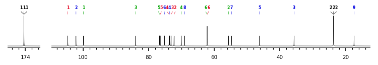
13C NMR data:
Linkage	Residue	C1	C2	C3	C4	C5	C6	C7	C8	C9
3,4,5	Ac	174.1-174.8	23.4-23.9
3,4,7	Ac	174.1-174.8	23.4-23.9
3,4	bXPsep	?	102.2	35.7	73.6	46.2	75.2	54.8	69.1	17.4
3	bDGalp	104.7	72.3	73.2	73.8	76.5	62.2
2	Ac	174.1-174.8	23.4-23.9
	bDGlcpN	99.9	55.7	84.0	70.1	76.7	62.2


1H NMR data:
Linkage	Residue	H1	H2	H3	H4	H5	H6	H7	H8	H9
3,4,5	Ac	-	1.94-1.99
3,4,7	Ac	-	1.94-1.99
3,4	bXPsep	-	-	1.80-2.60	4.06	4.31	3.69	4.03	4.14	1.13
3	bDGalp	4.43	3.52	3.62	4.47	3.68	3.65-3.68
2	Ac	-	1.94-1.99
	bDGlcpN	4.69	3.78	3.76	3.53	3.46	3.75-3.90


1H/13C HSQC data:
Linkage	Residue	C1/H1	C2/H2	C3/H3	C4/H4	C5/H5	C6/H6	C7/H7	C8/H8	C9/H9
3,4,5	Ac		23.4-23.9/1.94-1.99
3,4,7	Ac		23.4-23.9/1.94-1.99
3,4	bXPsep			35.7/1.80-2.60	73.6/4.06	46.2/4.31	75.2/3.69	54.8/4.03	69.1/4.14	17.4/1.13
3	bDGalp	104.7/4.43	72.3/3.52	73.2/3.62	73.8/4.47	76.5/3.68	62.2/3.65-3.68
2	Ac		23.4-23.9/1.94-1.99
	bDGlcpN	99.9/4.69	55.7/3.78	84.0/3.76	70.1/3.53	76.7/3.46	62.2/3.75-3.90
1H NMR data:
| Linkage | Residue | H1 | H2 | H3 | H4 | H5 | H6 | H7 | H8 | H9 |
|---|
| 3,4,5 | Ac |
| 1.94
1.99 | |
| 3,4,7 | Ac |
| 1.94
1.99 | |
| 3,4 | bXPsep |
|
| 1.80
2.60 | 4.06 | 4.31 | 3.69 | 4.03 | 4.14 | 1.13 |
| 3 | bDGalp | 4.43 | 3.52 | 3.62 | 4.47 | 3.68 | 3.65
3.68 | |
| 2 | Ac |
| 1.94
1.99 | |
| | bDGlcpN | 4.69 | 3.78 | 3.76 | 3.53 | 3.46 | 3.75
3.90 | |
|
13C NMR data:
| Linkage | Residue | C1 | C2 | C3 | C4 | C5 | C6 | C7 | C8 | C9 |
|---|
| 3,4,5 | Ac | 174.1
174.8 | 23.4
23.9 | |
| 3,4,7 | Ac | 174.1
174.8 | 23.4
23.9 | |
| 3,4 | bXPsep | ? | 102.2 | 35.7 | 73.6 | 46.2 | 75.2 | 54.8 | 69.1 | 17.4 |
| 3 | bDGalp | 104.7 | 72.3 | 73.2 | 73.8 | 76.5 | 62.2 | |
| 2 | Ac | 174.1
174.8 | 23.4
23.9 | |
| | bDGlcpN | 99.9 | 55.7 | 84.0 | 70.1 | 76.7 | 62.2 | |
|
The spectrum also has 1 signal at unknown position (not plotted). |
There is only one chemically distinct structure:
 report error
 Found 1 record.
Displayed record 1
Found 1 record.
Displayed record 1
