Taxonomic group: bacteria / Fusobacteria
(Phylum: Fusobacteria)
Host organism: Homo sapiens
Associated disease: periodontitis [ICD11:
DA0C 
];
infection due to Fusobacterium nucleatum [ICD11:
XN4P8 ]
The structure was elucidated in this paperNCBI PubMed ID: 28199859Publication DOI: 10.1016/j.carres.2017.01.009Journal NLM ID: 0043535Publisher: Elsevier
Correspondence: evguenii.vinogradov

nrc-cnrc.gc.ca
Institutions: Vaccine Program, Human Health Therapeutics Portfolio, National Research Council, Ottawa, ON, K1A 0R6, Canada, Department of Oral Biology, University at Buffalo, Buffalo, NY, 14214, USA
Fusobacterium nucleatum is an anaerobic bacterium found in the human mouth where it causes periodontitis. Recently, it has been gaining attention as a potential causative agent for colorectal cancer and is strongly linked with pregnancy complications including pre-term and still births. Little is known about virulence factors of this organism and thus we have initiated studies to examine the bacterial surface glycochemistry. Consistent with a recent paper suggesting that F. nucleatum strain 10593 can synthesize sialic acid, a staining technique identified sialic acid on the bacterial surface. We isolated lipopolysaccharide from this F. nucleatum strain and performed structural analysis on the O-antigen. Our studies identified a trisaccharide repeating unit of the O-antigen with the following structure: -[→4)-α-Neup5Ac-(2→4)-β-d-Galp-(1→3)-α-d-FucpNAc4NAc-(1-]- where Ac indicates 4-N-acetylation of approximately 30% FucNAc4N residues. The presence of sialic acid as a constituent of the O-antigen is consistent with recent data identifying de novo sialic acid synthesis in this strain.
Lipopolysaccharide, sialic acid, Fusobacterium, Fusobacterium nucleatum
Structure type: polymer chemical repeating unit
Location inside paper: abstract, p.39, table 1
Compound class: O-polysaccharide, O-antigen
Contained glycoepitopes: IEDB_136044,IEDB_136794,IEDB_137472,IEDB_141794,IEDB_142345,IEDB_146100,IEDB_149174,IEDB_190606,SB_165,SB_166,SB_170,SB_171,SB_172,SB_187,SB_195,SB_7,SB_84,SB_88
Methods: 13C NMR, 1H NMR, NMR-2D, GC-MS, sugar analysis, acid hydrolysis, MS/MS, methanolysis, GPC, reduction with NaBD4, acetylation, sialic acid staining
Comments, role: the NMR solution was not indicated; NMR data for aFucpNAc4N residue, 1H: 5.03 4.23 4.11 3.93 4.17 1.31, 13C: 95.4 48.8 75.8 56.3 64.1 16.8
NCBI Taxonomy refs (TaxIDs): 851Reference(s) to other database(s): GTC:G49573DR
Show glycosyltransferases
NMR conditions: at 298 K
[as TSV]
13C NMR data:
Linkage	Residue	C1	C2	C3	C4	C5	C6	C7	C8	C9
3,4,5	Ac	?	23.3
3,4	aXNeup	173.8	101.9	37.6	75.7	50.4	74.0	69.0	73.0	63.9
3	bDGalp	106.2	72.2	72.9	73.4	76.3	62.1
2	Ac	?	23.3
4	30%Ac	?	23.3
	aDFucpN4N	95.4	49.6	77.5	54.4	67.3	16.8


1H NMR data:
Linkage	Residue	H1	H2	H3	H4	H5	H6	H7	H8	H9
3,4,5	Ac	-	2.02-2.06
3,4	aXNeup	-	-	1.77-2.82	3.69	4.04	3.71	3.62	3.85	3.63-3.86
3	bDGalp	4.50	3.52	3.60	4.49	3.70	3.64-3.73
2	Ac	-	2.02-2.06
4	30%Ac	-	2.02-2.06
	aDFucpN4N	5.00	4.33	3.97	4.47	4.06	1.11


1H/13C HSQC data:
Linkage	Residue	C1/H1	C2/H2	C3/H3	C4/H4	C5/H5	C6/H6	C7/H7	C8/H8	C9/H9
3,4,5	Ac		23.3/2.02-2.06
3,4	aXNeup			37.6/1.77-2.82	75.7/3.69	50.4/4.04	74.0/3.71	69.0/3.62	73.0/3.85	63.9/3.63-3.86
3	bDGalp	106.2/4.50	72.2/3.52	72.9/3.60	73.4/4.49	76.3/3.70	62.1/3.64-3.73
2	Ac		23.3/2.02-2.06
4	30%Ac		23.3/2.02-2.06
	aDFucpN4N	95.4/5.00	49.6/4.33	77.5/3.97	54.4/4.47	67.3/4.06	16.8/1.11
1H NMR data:
| Linkage | Residue | H1 | H2 | H3 | H4 | H5 | H6 | H7 | H8 | H9 |
|---|
| 3,4,5 | Ac |
| 2.02
2.06 | |
| 3,4 | aXNeup |
|
| 1.77
2.82 | 3.69 | 4.04 | 3.71 | 3.62 | 3.85 | 3.63
3.86 |
| 3 | bDGalp | 4.50 | 3.52 | 3.60 | 4.49 | 3.70 | 3.64
3.73 | |
| 2 | Ac |
| 2.02
2.06 | |
| 4 | 30%Ac |
| 2.02
2.06 | |
| | aDFucpN4N | 5.00 | 4.33 | 3.97 | 4.47 | 4.06 | 1.11 | |
|
13C NMR data:
| Linkage | Residue | C1 | C2 | C3 | C4 | C5 | C6 | C7 | C8 | C9 |
|---|
| 3,4,5 | Ac | ? | 23.3 | |
| 3,4 | aXNeup | 173.8 | 101.9 | 37.6 | 75.7 | 50.4 | 74.0 | 69.0 | 73.0 | 63.9 |
| 3 | bDGalp | 106.2 | 72.2 | 72.9 | 73.4 | 76.3 | 62.1 | |
| 2 | Ac | ? | 23.3 | |
| 4 | 30%Ac | ? | 23.3 | |
| | aDFucpN4N | 95.4 | 49.6 | 77.5 | 54.4 | 67.3 | 16.8 | |
|
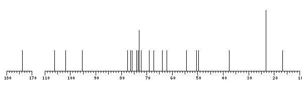
The spectrum also has 3 signals at unknown positions (not plotted). |
There is only one chemically distinct structure:
 report error
 Found 1 record.
Displayed record 1
Found 1 record.
Displayed record 1
