Taxonomic group: plant / Streptophyta
(Phylum: Streptophyta)
Organ / tissue: fruit
The structure was elucidated in this paperNCBI PubMed ID: 10606546Publication DOI: 10.1021/jf990608vJournal NLM ID: 0374755Publisher: American Chemical Society
Correspondence: Ho CT <ho

aesop.rutgers.edu>
Institutions: Department of Food Science and Center for Advanced Food Technology, Rutgers University, 65 Dudley Road, New Brunswick, New Jersey 08901-8520, USA, Department of Food and Nutrition, Osaka City University, 3-3-138 Sugimoto Sumiyoshi, Osaka, Japan, Pacific Biomedical Research Center, University of Hawaii, 1993 East-West Road, Honolulu, Hawaii 96822, USA
Two known glycosides and a novel trisaccharide fatty acid ester were isolated from the n-butanolsoluble fraction of the fruits of Morinda citrifolia (Noni). Structure determination was carried out by spectral techniques such as MS, IR, NMR, and 2D-NMR. The novel trisaccharide fatty acid ester was elucidated as 2,6-di-O-(β-D-glucopyranosyl)-1-O-octanoyl-β-D-glucopyranose. The known compounds were identified as rutin and asperulosidic acid.
2, rutin, Noni, Morinda citrifolia, trisaccharide fatty acid ester, 6-di-O-(β-D-glucopyranosyl-1-O-octanoyl-β-D-glucopyranose, asperulosidic acid
Structure type: oligomer ; 609 [M-H]-
C
27H
30O
16Location inside paper: compound 2, fig. 1(2)
Trivial name: rutin
Compound class: glycoside, flavonoid glycoside, flavone glycoside
Contained glycoepitopes: IEDB_136105,IEDB_142488,IEDB_144144,IEDB_146664,IEDB_225177,IEDB_885823,IEDB_983931,SB_192
Methods: 13C NMR, 1H NMR, IR, FAB-MS, HMBC, HMQC, COSY, NOESY, APCI-MS
Comments, role: NMR temperature was not specified
Related record ID(s): 63269, 63271
NCBI Taxonomy refs (TaxIDs): 43522
Show glycosyltransferases
NMR conditions: in DMSO-d6
[as TSV]
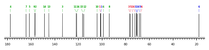
13C NMR data:
Linkage	Residue	C1	C2	C3	C4	C5	C6	C7	C8	C9	C10	C11	C12	C13	C14	C15	C16
3,6	aLRhap	101.0	70.6	70.8	70.3	68.5	18.0
3	bDGlcp	101.4	74.3	76.7	72.1	76.1	67.3
	xXQuercetin	-	156.7	133.5	177.6	161.5	98.9	164.3	93.9	156.9	104.2	121.9	115.5	145.0	148.7	116.5	121.4


1H NMR data:
Linkage	Residue	H1	H2	H3	H4	H5	H6	H7	H8	H9	H10	H11	H12	H13	H14	H15	H16
3,6	aLRhap	4.40	3.00-3.84	3.00-3.84	3.00-3.84	3.00-3.84	1.00
3	bDGlcp	5.35	3.00-3.84	3.00-3.84	3.00-3.84	3.00-3.84	3.00-3.84
	xXQuercetin	-	-	-	-	-	6.22	-	6.41	-	-	-	7.56	-	-	6.86	7.56


1H/13C HSQC data:
Linkage	Residue	C1/H1	C2/H2	C3/H3	C4/H4	C5/H5	C6/H6	C7/H7	C8/H8	C9/H9	C10/H10	C11/H11	C12/H12	C13/H13	C14/H14	C15/H15	C16/H16
3,6	aLRhap	101.0/4.40	70.6/3.00-3.84	70.8/3.00-3.84	70.3/3.00-3.84	68.5/3.00-3.84	18.0/1.00
3	bDGlcp	101.4/5.35	74.3/3.00-3.84	76.7/3.00-3.84	72.1/3.00-3.84	76.1/3.00-3.84	67.3/3.00-3.84
	xXQuercetin						98.9/6.22		93.9/6.41				115.5/7.56			116.5/6.86	121.4/7.56
1H NMR data:
| Linkage | Residue | H1 | H2 | H3 | H4 | H5 | H6 | H7 | H8 | H9 | H10 | H11 | H12 | H13 | H14 | H15 | H16 |
|---|
| 3,6 | aLRhap | 4.40 | 3.00
3.84 | 3.00
3.84 | 3.00
3.84 | 3.00
3.84 | 1.00 | |
| 3 | bDGlcp | 5.35 | 3.00
3.84 | 3.00
3.84 | 3.00
3.84 | 3.00
3.84 | 3.00
3.84 | |
| | xXQuercetin |
|
|
|
|
| 6.22 |
| 6.41 |
|
|
| 7.56 |
|
| 6.86 | 7.56 |
|
13C NMR data:
| Linkage | Residue | C1 | C2 | C3 | C4 | C5 | C6 | C7 | C8 | C9 | C10 | C11 | C12 | C13 | C14 | C15 | C16 |
|---|
| 3,6 | aLRhap | 101.0 | 70.6 | 70.8 | 70.3 | 68.5 | 18.0 | |
| 3 | bDGlcp | 101.4 | 74.3 | 76.7 | 72.1 | 76.1 | 67.3 | |
| | xXQuercetin |
| 156.7 | 133.5 | 177.6 | 161.5 | 98.9 | 164.3 | 93.9 | 156.9 | 104.2 | 121.9 | 115.5 | 145.0 | 148.7 | 116.5 | 121.4 |
|
The spectrum also has 1 signal at unknown position (not plotted). |
There is only one chemically distinct structure:
 Found
Found  report error