Taxonomic group: bacteria / Proteobacteria
(Phylum: Proteobacteria)
Associated disease: infection due to Escherichia coli [ICD11:
XN6P4 
]
The structure was elucidated in this paperNCBI PubMed ID: 7544238Publication DOI: 10.1016/0008-6215(95)00041-QJournal NLM ID: 0043535Publisher: Elsevier
Correspondence: torgov

ioc.ac.ru
Institutions: Max-Planck-Institut für Immunbiologie, Freiburg, Germany
Structures for the N-acetylneuraminic acid (Neu5Ac)-containing O56 and O24 polysaccharides of Escherichia coli have been reported previously. During these studies unusual chemical shifts had been observed for the NMR signals for H-3eq and C-3 of the Neu5Ac residues of both polysaccharides. In further pursuing this phenomenon, we have reinvestigated the O56 and O24 polysaccharides as well as derived oligosaccharides by one- and two-dimensional NMR spectroscopy. The results showed that structures of both polysaccharides (PSs) had to be modified and formulated as [formula: see text] 2D ROESY spectra revealed a strong NOE between H-3eq of Neu5Ac and the protons of the side-chain sugar (H-3 and H-5 of α-D-Galp in the O56 PS and H-3 of α-D-Glcp in the O24 PS) and also between H-3ax of Neu5Ac and H-3 of β-D-Glcp in the main chain. This indicated a close spatial association of the seven-linked α-Neu5Ac and the side-chain residues α-D-Galp (O56 PS) and α-D-Glcp (O25 PS), respectively. The strong long-range spatial contacts caused the unusual chemical shifts of H-3eq and C-3 of Neu5Ac.
Escherichia coli, NMR spectroscopy, polysaccharide structure, 024 and 056 antigens
Structure type: suggested polymer biological repeating unit
Location inside paper: abstract, structure V, Table 8
Compound class: O-polysaccharide, O-antigen
Contained glycoepitopes: IEDB_135813,IEDB_136794,IEDB_136906,IEDB_137340,IEDB_137472,IEDB_141794,IEDB_141807,IEDB_142488,IEDB_146100,IEDB_146664,IEDB_149174,IEDB_151528,IEDB_151531,IEDB_190606,IEDB_983931,SB_170,SB_171,SB_172,SB_192,SB_7,SB_84
Methods: NMR-2D, NMR, sugar analysis, Smith degradation
Comments, role: revision of structure ID 117052; chemical repeat frame is different in the paper; carbon 5 in aXNeup at #3,3 has a +15.4 ppm mismatch in 13C spectra (experiment: 69.6; hybrid simulation in water: 54.2, trust 60%)
Related record ID(s): 326, 327, 328, 8382, 8411, 8412, 8413, 8414, 20438, 20659, 22619, 25691, 108697, 124618
NCBI Taxonomy refs (TaxIDs): 562Reference(s) to other database(s): GTC:G32692RT, GlycomeDB:
6888
Show glycosyltransferases
NMR conditions: in D2O
[as TSV]
13C NMR data:
Linkage	Residue	C1	C2	C3	C4	C5	C6	C7	C8	C9
3,3,5	Ac
3,3	aXNeup	173.9	102.4	37.75	69.6	?	72.5	78.9	73.0	63.7
3,2	aDGalp	97.2	69.6	70.6	70.5	71.55	62.4
3	bDGlcp	103.3	73.6	78.7	69.9	76.75	62.4
2	Ac
	bDGlcpN	102.2	57.3	80.8	70.6	76.3	62.45


1H NMR data:
Linkage	Residue	H1	H2	H3	H4	H5	H6	H7	H8	H9
3,3,5	Ac
3,3	aXNeup	-	-	2.0-2.51	4.05	3.91	3.88	3.72	3.78	3.50-3.80
3,2	aDGalp	5.52	3.83	3.92	4.00	4.13	3.75
3	bDGlcp	4.70	3.66	4.15	3.54	3.50	3.73-3.90
2	Ac
	bDGlcpN	4.74	3.68	4.08	3.52	3.53	3.79-4.00


1H/13C HSQC data:
Linkage	Residue	C1/H1	C2/H2	C3/H3	C4/H4	C5/H5	C6/H6	C7/H7	C8/H8	C9/H9
3,3,5	Ac
3,3	aXNeup			37.75/2.0-2.51	69.6/4.05	?/3.91	72.5/3.88	78.9/3.72	73.0/3.78	63.7/3.50-3.80
3,2	aDGalp	97.2/5.52	69.6/3.83	70.6/3.92	70.5/4.00	71.55/4.13	62.4/3.75
3	bDGlcp	103.3/4.70	73.6/3.66	78.7/4.15	69.9/3.54	76.75/3.50	62.4/3.73-3.90
2	Ac
	bDGlcpN	102.2/4.74	57.3/3.68	80.8/4.08	70.6/3.52	76.3/3.53	62.45/3.79-4.00
1H NMR data:
| Linkage | Residue | H1 | H2 | H3 | H4 | H5 | H6 | H7 | H8 | H9 |
|---|
| 3,3,5 | Ac | |
| 3,3 | aXNeup |
|
| 2.0
2.51 | 4.05 | 3.91 | 3.88 | 3.72 | 3.78 | 3.50
3.80 |
| 3,2 | aDGalp | 5.52 | 3.83 | 3.92 | 4.00 | 4.13 | 3.75 | |
| 3 | bDGlcp | 4.70 | 3.66 | 4.15 | 3.54 | 3.50 | 3.73
3.90 | |
| 2 | Ac | |
| | bDGlcpN | 4.74 | 3.68 | 4.08 | 3.52 | 3.53 | 3.79
4.00 | |
|
13C NMR data:
| Linkage | Residue | C1 | C2 | C3 | C4 | C5 | C6 | C7 | C8 | C9 |
|---|
| 3,3,5 | Ac | |
| 3,3 | aXNeup | 173.9 | 102.4 | 37.75 | 69.6 | ? | 72.5 | 78.9 | 73.0 | 63.7 |
| 3,2 | aDGalp | 97.2 | 69.6 | 70.6 | 70.5 | 71.55 | 62.4 | |
| 3 | bDGlcp | 103.3 | 73.6 | 78.7 | 69.9 | 76.75 | 62.4 | |
| 2 | Ac | |
| | bDGlcpN | 102.2 | 57.3 | 80.8 | 70.6 | 76.3 | 62.45 | |
|
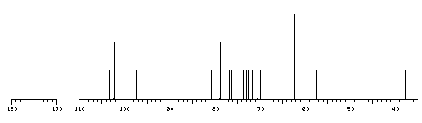
The spectrum also has 1 signal at unknown position (not plotted). |
There is only one chemically distinct structure:
 report error
 Found 1 record.
Displayed record 1
Found 1 record.
Displayed record 1
